So before I get into this post let me begin with a minor disclaimer: Neither Michael and I are "Artistically Gifted" so our poster will
NOT be featured in this post.
So basically last week we got the assignment of drawing a poster on the "Fluid Mosaic Cell Membrane Model". Sounds like fun right? Surprisingly.............it was, despite our not using markers and any artistic ability since third grade.
 |
| Mosaic Model (not our poster) |
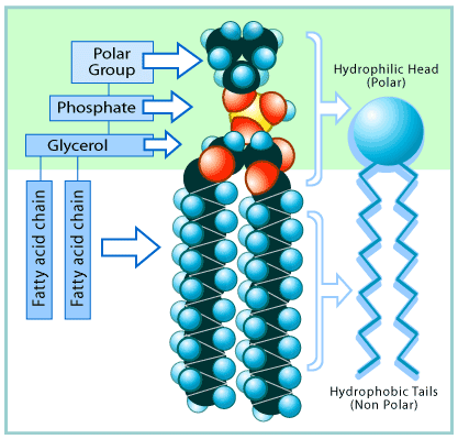
The cell membrane (outside of the cell (works like skin)) is used to control what goes in and out of a cell, ie: food and waste. It is accurately described as a "Fluid Mosaic" because, like glass mosaic it is made up of
LOTS of little pieces and like a fluid it is flexible, moving as needed without breaking. The main part of the cell boundary (the "wall" that keeps "stuff" out) is made up of Phospholipids (A hydrophilic phosphorus head (the little circles) attached onto two fatty (lipid) tails). Because the heads are hydrophilic (they like water) they position themselves on the edges of the cell membrane, the hydrophobic ( water fearing- in latin) tails go to the inside of the membrane because they are
VERY scared of water. The head of the phospholipid has polar group on the top which forms the bonds that hold mosaic together (from head to head), giving it fluidity.
 |
| A Magical Phospholipid! |
Now, do you see those big blue lumbering blobs that are interrupting the beautiful mosaic of phospholipids are proteins, and yes, they serve a function (amazing right?). The middle protein in the picture (the one that looks like it has horns) is called a receptor protein. Receptor proteins are a type of integral protein (proteins integrated into the membrane) that is used for signaling between cells. The receptor proteins absorb the signal molecules sent out by other cells (like in the nerve system) that tell the receiving cell to "fire". The little valley on the top of that protein is where the molecules "dock". Way out in the back of the protein (back in the middle of nowhere, like La Junta) is a glycoprotein. Glycoproteins are polypeptides (short polymers of amino acids linked by peptide bonds) with a Glycan (the carb structure sticking out) attached. Glycoproteins are used for cell to cell interactions. On the left side of the image there is a recognition protein. Recognition proteins (like their name suggests) are used by cells to identify that they belong in the body. When a person gets an organ transplant the recognition proteins are different so the body will sometimes attack the new organ because it is viewed as a foreign and possibly hostile object.
The last type of protein in the cell membrane is called a transport protein. This protein is possibly the most important protein in the entire membrane (at least in my opinion). Since the membrane (also called the lipid bilayer), is basically a giant
impenetrable wall that surounds the yummy goodness that is the internal components of the cell, there needs to be a way to get things in (food, vital molecules) and let stuff (Waste, excess molecules) out. Thus, the transport protein was born. All that a transport protein is simply put, is a doorway into a cell. There is one minor condition though to transport proteins, each one only lets in a certain type of molecule. If I was to use a metaphor to describe it I would compare them to bathroom doors. Only guys go into the men's bathroom and only girls go into the women's bathroom (hopefully). If one sex was to go through the wrong door (very rare) bad things would happen (think pepper spray and screaming. followed by sexual harassment charges....) Since we have determined that there are specific transport proteins for the different types of molecules (sodium and the like) we now have to think about the two types of transport proteins, defined by how they work.
 |
| Transport Proteins. |
The first type of transport proteins are channel proteins. Channel proteins span the entire width of the lipid bilayer and use passive transport to move molecules. Passive transport uses no chemical energy to move molecules so it is limited by the size of the molecule. Passive transport is also limited by the chemical gradient (molecules move from high to low concentration). On a cell membrane this is known as facilitated diffusion.
 |
| Facilitated Diffusion |
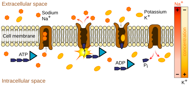
The second type of transport proteins are Carrier proteins. Carrier proteins use chemical energy to move molecules against the concentration gradient and molecules that are to large for a protein channel. Carrier proteins are used to move vital molecules against the concentration gradient so that the cell can survive. If it uses adenosine triphosphate (ATP) as an energy source then it is termed Primary Active Transport. If it uses an electrochemical gradient the it is Secondary Active Transport.
 |
| Primary Active Transport Using ATP |
Since transport proteins are necessary for getting molecules in and out of a cell, when something goes wrong with them it can result in pretty nasty disease. Cystic Fibrosis (CF) is one of those diseases. In the cells of people with CF the epithelial cells in the lungs are poorly permeable to Chloride ions (Cl-). Damaged transport proteins are responsible for this.
 |
| Epithelial Cells |
The lining cells have channels on their outside surface (on the side of the airway). One of the channels allows sodium ions to flow into the cell and the other controls the passage of chloride ions out of the cell into the mucus on the airway surface. Along with the ion pump, the action of the channels results in an excess of chloride ions in the mucus; an ionic gradient is set up, with a higher concentration towards the outside. In an attempt to equalize the salt concentrations, water is dragged out through the gaps between the cells and this keeps the mucus moist. If a person has CF then the all important chloride channel is blocked. This means that there is no movement of chloride ions into the mucus. With no ionic gradient, there is no need for water to move towards the surface and the mucus dries out. In 1989 the defective gene for CF was isolated (kinda). There are over 800 different mutations of the gene, with each one affecting the correct function of the Chloride Channel (CF transmembrane regulator (CTFR)) According to <http://resources.schoolscience.co.uk/MRC/3/page3.html> it was 'one of the most significant discoveries in the history of human genetics'. It led directly to improved diagnosis of the disorder and has improved the genetic counseling offered to affected families.
In conclusion, Holy Crap. These very tiny little cells that make up our body are super complicated and very organized. If one little piece of the cells go bad then your whole body gets screwed up. Its like a swiss watch (had enough of my metaphors yet?) if one little gear is missing it all goes to hell.


No comments:
Post a Comment