Search This Blog

Showing posts with label Graded. Show all posts

Wednesday, March 30, 2011

The ABC's of DNA sequencing...(ish)

Want to hear a (anti) joke?   Ok, here goes: "Abby, Bob, and Carol walk into a hospital.......and get their DNA tested for a horrible disease." Because the hospital that they walked into a hospital stuck in the 1980's they are using the Sanger method of sequencing to examine the DNA. To make a complex process rather simple, the Sanger method takes a sample of DNA from a subject, each base (A,T,G, C) is radioactively labeled (for detection later in a sequencing machine) and synthesized (copied). The newly synthesized DNA is then put through gel electrophoresis, which uses electricity and gel to sort the DNA Molecules by size and creates a visual representation on X-Ray fil, or the gel itself. (Figure 1)

(Fig. 1)
So, Abby, Bob, and Carol all had their DNA run through the Sanger process. The next step to get their results was for a "skilled" technician to write out the DNA sequence out. To write out the sequence you start at the bottom and write down the the letter that each mark represents. (the above sequence would be CGA GAT ATA etc...) Each letter represents a base, and he three letter sorting is important later on to determine the proteins that the sequence represents.

Abby's Sequence went:  ATG GTG CAC CTG ACT CCT GTG GAG AAG TCT GCC

Bob's Sequence went:    ATG GTG CAC CTG ACT CCT GTG GAG TAG TCT GCC

Carol's Sequence went:  ATG GTG CAC CTG ACC CTG AGG AGA AGT CTG CCC

In order to determine if they had the "life ending disease" their DNA sequences were compared to that a control subject (Norm) who did not have the disease.

Norm's Sequence was:   ATG GTG CAC CTG ACT CCT GAG GAG AAG TCT GCC

To determine the likely hood of having the disease the hospital needs to create a quantitative representation of what all those letters mean, so the create a number called the percent similarity. To do this the put the number of bases that are simular to "Norms" over the total number of bases in the sequence. I took the liberty of putting the results into a pretty little graph to show how each of the patients compares to the control (Norm.).


As you can very clearly see, Bob and Abby have a very simular (96.45%) DNA sequence to the control, which would lead me to assume that they would not have the horrible disease. Carol on the other hand was not that simular to Norm, therefore I would assume that she has the disease. 

One Last Note: (for content stuffs) 
The reason that DNA is sequenced in chunks of three (called codons) because the proteins that are formed based on the DNA are created from the three letter sequences. Each set of three letters, in other words, represents a protein. To determine what proteins are made by what sequence, one has to look at a magical "decoder table." The nice thing about proteins and DNA sequences, is that the same protein can usually be made a number of different sequences, so long as the first and second letters of the sequence are the same. For example, the sequences CCU CCC CCA and CCG all make "pro" (Proline.) Which means that if a sequence is off, the proteins won't always be off. Which is the important part...

Wednesday, March 16, 2011

Eugenics. Post (3 Pages? Psh....)

So, this week (ok maybe six weeks ago) we started a project on Eugenics (which was a minor factor in the delay of my last blog post). Eugenics is "the applied science or biosocial movement which advocates the use of practices aimed at improving the genetic composition of a population," at least according to wikipedia. In much simpler terms it's all about controlling the population's genetic makeup in order to ensure a "perfect" population. So, Lets look at the essential questions of this rather unsavory "biological science":


What were the social origins of eugenics?
 Eugenics developed in the wake of economic and social problems following the civil war. Depressions occurred every decade from 1876 to the beginnings of the 20th century. These depressions fueled a labor unrest, caused by militant labor unions and the rapid growth of american industry. Social Darwinism had attempted to explain the inequalities experienced by the lower class as "survival of the fittest", but by the the beginning of th 20th century that idea had been flipped.  Those people who sat at the top of the economy in the upper class had a declining birthrate. Meaning that they were in fact losing the struggle for existence. Not only were the working  class working and organizing against them, but they were also out-reproducing them. To solve all the new problems that came with industrialization, a new philosophy called progressivism emerged. Progressivism was all about managed capitalism- letting the government and planning have an increased role in the market. The biggest component of this was the idea of scientific management: long-term planning by specially trained university experts. This new "managerial" class would soon become important to the managerial process, science was seen as the new "cure-all" that would bring in a new era of control to nature and human society. Suddenly, the the idea that genetics explained the underlined causes of human social problems (feeble-mindedness, alcoholism, etc...) came about. These issues were said to be caused by "the inheritance of defective germ plasm", believers argued that society paid a very high price by allowing those people (who would eventually be cared for by the state) to be born. Their solution? Sterilize that "defective" adult. By doing this, eugenicists claimed that future generations could save thousands of dollars. Another problem that eugenics solved was the mutual antipathy, shared by eugenicists and their wealthy supporters, towards militant labor unions and political revolution. Both of which were seen to be caused by the large influx of immigrants from southern Europe. Eugenicists then claimed to have data that showed the problem was in their genes, effectively placing the blame in "germ plasm" and ethnic groups, rather than in the structure of society its self. In essence, they managed to use science as an excuse to blame victims for their own problems. Basically, eugenics offered the prospect of a smooth transition to a better future, and a method to attack social problems at their roots.

What were the scientific origins of eugenics?
In 1883 Francis Galton coined the term eugenics as a moral philosophy to encourage the healthiest, and most able people to have more children. His version is known as "positive eugenics". Negative eugenics supported the removal of the weakest members from a population. Needless to say the negative version was favored more...The idea of segregating people deemed unfit to reproduce dates back to antiquity. Concerns about environmental influences that could damage heredity were formalized in the 1700's as "degeneracy theory". It would maintain a strong following up until the 19th century. Onanism was presented as the first cause of degeneracy in medical schools. This fear of degeneracy through onanism prompted prison doctor Henry Clay Sharp to carry out vasectomies on prisoners beginning in 1899, later resulting a law that would make it mandatory in Indiana. Benedict Morel's work extended the causes of degeneracy to include poisoning by toxic substances. Richard Dugale believed that  good environments could turn a degenerate into a worthy citizen through three generations. He conducted a study (The Jukes, 1877) to prove this. In the 1880's August Wiseman's theory on germ plasm managed to convince most people that changes to the body tissues (the soma) had no effect on the reproductive tissues (the germ plasm). So, naturally this theory was favored by negative eugenics. Go figure..  Most basic scientists would shun eugenics as vulgar, but their contributions to the field of genetics would be absorbed by eugenicists. Eventually, Mendelian analysis would be part of most eugenicists backgrounds, as would agriculture where most eugenicists were present [like the american breeders ass. (ABA)]. Evolutionary models would also contribute to eugenic theory, with fears that the smarter families would have less kids than the degenerate families due to welfare supporting these people, which upset the role of natural selection. Soon even medicine hopped on the bandwagon, with doctors advocating medicine and programs to deal with degenerates. Most would argue that sterilization was the best plan for dealing with degenerates. With vasectomies and tubal litigation as the preferred methods because they did not alter the physical or psychological contributions of reproductive organs as well as saving the state money by not having to institutionalize them.

What research methods were used to study eugenics and what were their flaws?
At the core of eugenics was the was a research effort to apply Mendel's laws to the inheritance of human traits. Eugenicists examined family pedigrees to determine the inheritance pattern of a trait, and then score family members for the presence or absence of that trait. They succeeded quite well on the first half, as large families were easy to spot patterns in, but scoring members was especially difficult due to the complexity of most traits. The main problem here was that researchers were lax in defining the traits they were studying. Most of the traits (mental and behavioral) they were looking for were subjective, so the diagnosis was often mishandled, corrupting their data. The second issue with their research was that they often treated large complex issues as if they were caused by one thing, and not multiple variables combining into one issue. Data was also affected by poor survey and statistical methods. Most researchers got their information from second had sources and heresy, due to to a lack of good medical records. Also, when presenting results, eugenicists were VERY biased, using their data to falsely exaggerate claims. False quantification also plagued researchers. For example IQ tests that were dependent on cultural knowledge were given to immigrants under a wide variety of conditions, and were counted as official evidence that immigrants were feeble minded.  To simplify it all, the research that was done was biased, not properly administrated, and to many variables were left open.

How did eugenics research impact American society?
Eugenics had a very large negative impact on American society. In a period of changing times, marked by industrial revolutions, and an influx of immigration. Eugenics promoted racial in equality, social inequality, and a sense of hatred against the lower class in America. Numerous rights of people were violated with the passing of marriage laws (preventing people from marrying outside their race), sterilization laws (forcibly sterilizing those with "bad" germ plasm), and the immigration restriction that would result from studies showing that they were bad for the population. The affects of American eugenics could also be felt across the world, with German leaders (on trial for war crimes Nuremberg) citing American eugenicists as the inspiration behind Hitlers purification laws, and the eventual genocide of the Holocaust.

DNA-My Model, and an gentle explanation

Recently in Biology we have been working on a rather fun concept: DeoxyRibonucleic Acid otherwise known as DNA. So, I made a model of it on Google Docs (*cough* tech savvy 4 *cough*) in order to study for the DNA quiz last week.  So here goes my explanation of DNA's structure, and what it all means...


Double Helix (Fig. 1)
At the base of DNA are little cornerstones known as nucleotides. A nucleotide is made up of three pieces a phosphate (the white circle), a five carbon sugar (deoxyribose, the black pentagon) and a nitrogenous base (one of the colorful weird pieces in the middle). Nucleotides first join together by connecting together the front pieces sugar with the second pieces phosphate creating a single chain. The second place that nucleotides link together, to create a double chain (Kiel's brand new descriptive word of the day) is in the middle linking two nitrogenous bases together. The only condition with nitrogenous bases connecting together is that A (Adenine) bases can only link with T (Thymine) bases and C (Cytosine) bases must link with G (Guanine) bases. It is that pattern of bases (read three at a time) that are the genetic instructions used in the development functioning of all living organisms. The flat structure shown above, in real life, is twisted into a double helix (fig. 1) and get wrapped around a protein in the cell (but, thats way above the scope of this post). (fig. 2).
DNA wrapped around a DNA-Binding Protein (Fig. 2)


Wednesday, February 16, 2011

Meiosis, Mitosis, Reproduction...and everything else I've been to lazy to post about.

If you couldn't tell by the title of this post, I've been slacking quote bad with my biology posts (Damn you stumble upon.) These past 2 (3?) (4?) weeks we've been covering Meiosis, Mitosis, Stemcells, Cloning, Reproduction, and Genetics. Which means it's time for another series of blog posts to cover all the information. So the official topic of this post will be "How Cells Reproduce" and "How a baby is made (ish)."

How Cells Reproduce, and Make Babies. Its Magical.

Once upon a time, you were a cell. As a cell you couldn't move, breathe, talk or hear, but the one thing that you could do is reproduce, and reproduce you did. Starting as one cell and then doubling exponentially until you ended up as a body with 50-75 trillion cells. The processes that control how that original cell is made, and how a person is made from that cell are some of the most important and intricate process in biology.

Disclaimer: 
Now the first thing that I should explain is how the cell cycle and Mitosis work, as these will be necessary to understand how Meiosis (slightly more complicated than Mitosis) works, and how the baby duplicates its cells to grow in the womb. 

Mitosis & The Cell Cycle:
Since Mitosis is a circular cycle (seems redundant right?) Which means the new cycle starts right after the last cycle ended. Now that the last cell cycle has ended our newly created (cloned would be a more accurate term?) cell (let's call it Joe) enters the magical phase of G1 (not G6, G1, that song is just stupid). The G1 phase is all about...you guessed it, Growth! All that the cell does during G1 is get nice and big to prepare to duplicate its chromosomes. This happens in the next stage, aptly named Synthesis. All that happens in this stage is the chromosomes make clones of themselves going from 23 pairs of chromosomes (2 sets of 23) to 46 pairs (4 sets of 23.) From here Joe then enters phase G2, also known as.......wait for it.......wait for it.......wait......GROWTH PHASE TWO!!!!!!! During this phase the cell gets to a large enough size that it cannot efficiently remove waste from the insides, and get "food" in. Which, of course, means it's time to end interphase (the last three phases/stages) and enter...Duh Duh Duh......the Mitotic cycle!!!  

The Cell Cycle. Obviously.


This is where the action starts. The mitotic cycle consists of four separate phases: Prophase, Metaphase, Anaphase and Telophase. Prophase, the first phase, is when the chromosomes become visible (under a microscope) and the nucleolus magically dissolves. During this phase the pairs of chromosomes, due to their cloning, are in an "X" shape, where the original, and duplicate are crossed over each other. These pairs are joined to the cells by spindles attached to "Centromere's" 

"X" shape.

Then comes metaphase, where the chromosomes lined up across the center of the cell, across what as known as the metaphase plate, by the spindles attached to the centromeres. In anaphase this lining up pays off because, the pairs are then pulled apart by the centromeres to the opposite sides of the cells. In telophase the spindles disappear and the cell membrane starts to split apart into daughter cells.  When the cell enters the final stage of mitosis, and the cell cycle, cytokinesis, the cell splits into two daughter cells, each with 23 pairs of chromosomes. 
Mitosis-ness....In image form!


Meiosis & The Beginning of a Baby
When a mommy and a daddy love each other very much, (or 16 year old's want to get popular, yes I do hate MTV's programing) a baby is made. Ok, I'm joking. It's a lot more complicated than that. The process of cellular reproduction starts back when parents are just babies themselves. Creating a sort of never ending cycle of "which came first?" In order to create the cell that replicates into a baby, the parents must have sex cells (called gametes) to produce the sperm and the egg that join up and become a baby. Now, most people know that each cell has 46 chromosomes, organized into 23 pairs. But for a baby to be formed the sperm and egg have to have half that number (23 unpaired chromosomes) this happens through a process called meiosis. As Michael Rees, in his blog post "Cell's: They Reproduce." said:

  "First off, the goal of meiosis is to produce a cell with only twenty-three chromosomes so that it can share its chromosomes with another cell in order to produce a cell with unique genes that is then 
capable of developing into a baby of the species."  

Simply put, Meiosis is Mitosis with an extra division added on to the end, allowing you to get 23 individual chromosomes rather than the 23 pairs normally produced in Mitosis. Meiosis follow's the basic pattern of mitosis, but at the end, it adds in an extra division of the daughter cells. By adding that extra division at the end of the mitotic cycle,  the daughter cells of "Joe" do not get to go into synthesis, which then means that they don't double their number of chromosomes, which allows each daughter cell to get split into cells that each contain 23 individual chromosomes. Because sex cells each have 23 chromosomes they are able to combine into one cell with 23 pairs of chromosomes, which then divides by mitosis into more cells, which then create a baby. Simple right?


Friday, December 17, 2010

Photosynthesis&Respiration

So this blog post looks like its going to be a rather big project, getting two content standards to fit in one blog post....(sadly I'm doing this the night before grades are final...oh well) The aim of this post is to basically convey my understanding of photosynthesis, respiration, and how the energy produced is used. Unlike a number of my peers, rather than do the green human project, I decided, in the interest of time, just to do a flat out explanation of the two processes. So here goes.....

Photosynthesis. Or, How to make your own food.......

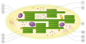
Essentially, photosynthesis is the process by which photoautotrophs synthesize food (sugar) from carbon dioxide (CO2) using the energy from light. Sounds mighty dang simple right? Well, it mostly is. It happens like this: In oxegenic photosynthesis (literally photosynthesis that releases oxygen) Light energy is absorbed by proteins that contain chloroplasts which contain chlorophylls (by the way, chlorophylls are green because that is the part of the light spectrum they do not use). The chlorophylls store most of the energy in the form of ATP.

Chloroplast

The next step in photosynthesis is the oxidation of H20. Energy (ADP) is used to split the hydrogen and Oxygen molecules apart. The oxygen molecules are released (they can be used in respiration), and the hydrogens are kept. When CO2 is added to the process they each lose an Oxygen, which attach to some of the hydrogen molecules, forming H2O. The remaining Hydrogen, Oxygen, and Carbon then all get combined into glucose.

---CO2 can also be converted into sugars suing a process called carbon fixation. The most common type of carbon fixation in biological life is the Calvin cycle. The Calvin cycle describes the way that light energy is used in the creation of chemical free energy, stored in glucose. The key enzyme that makes the cycle run is called RubBisCO, found in the chloroplast stroma. The Calvin cycle includes a number of regulatory functions that prevent it from being respired (look at the second half of the post) into CO2 (preventing energy (ATP) from being wasted without a net gain). For more information on the Calvin cycle, click here.-----

The glucose can then be burned through cellular respiration.

Cellular Respiration Or:how you use your food
Cellular respiration is the process by which nutrients are broken down into ADP. The first kind of respiration is called aerobic respiration. Aerobic respiration requires oxygen to break down the nutrients. It has three main stages. Glycosis, Krebs cycle, and electron transport.


  1. In Glycolysis (spliting sugars), Glucose, is split into two molecules of a three carbon sugar. In the process, two molecules of ATP, two molecules of pyruvic acid and two "high energy" electron carrying molecules of NADH are produced. Glycolysis can occur with or without oxygen. In the presence of oxygen (like in aerobic respiration), glycolysis is the first stage of cellular respiration. Without oxygen, glycolysis allows cells to make small amounts of ATP (fermentation).
  2.  The Krebs Cycle (citric acid cycle) begins after the two molecules of the three carbon sugar produced in glycolysis are converted to a different compound (acetyl CoA). Through a series of intermediate steps, several compounds capable of storing electrons are produced along with two ATP molecules. These compounds, known as NAD (nicotinamide adenine dinucleotide...what is it with bio and big words?) and flavin adenine dinucleotide (FAD), are reduced in the process. These reduced forms carry the electrons to the next stage. The Krebs Cycle occurs only when oxygen is present but it doesn't use oxygen directly (in aerobic respiration).   
  3.  Electron Transport requires oxygen directly. The electron transport "chain" is a series of electron carriers in the membrane of the mitochondria in eukaryotic cells. Through a series of reactions, the earlier mentioned electrons are passed to oxygen. In the process, a gradient is formed, and eventually ATP is produced.

Maximum ATP Yields: P
rokaryotic cells can yield a maximum of 38 ATP molecules while eukaryotic cells can yield a maximum of 36. In eukaryotic cells, the NADH molecules produced in glycolysis pass through the mitochondrial membrane, which "costs" two ATP molecules.


Other kinds of cellular respiration include fermentation, where the pyruvate is converted to waste products, which when removed from the cell oxidizes the electron carriers, and anaerobic respiration wher unlike in aerobic, the oxygen is replaced by an inorganic acceptor (sulfur) is used. 

Wednesday, December 15, 2010

Enzyme Lab. With graphs.

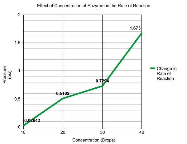
A few weeks ago (if you haven't noticed there's about a three week period between the time we do stuff in class and the day I write a post for it) we did what was called the "Enzyme Lab". This lab consisted of mixing 3ml of H2O2 (hydrogen peroxide) with 3ml of water (H20) , and adding an enzyme (yeast) to start a chemical reaction that produces O2. The way the experiment works is the enzyme breaks the Oxygen molecules off of the H2O and H2O2 and releases them. We measured the reaction with a pressure sensor that was hooked up to my mac (which sucks slightly less...). Of course no experiment would be complete without a few variables, so we did the experiment three different ways each time changing one aspect of it (we repeated each set three times in order to gain an accurate representation of data). The first time we changed the concentration of the enzyme (10 drops, 20 drops, 30 drops, 40 drops.), the second time we changed the temperature of the solution be for adding the enzyme (Hot, cold, warm, and room temp). The third time we substituted the water (H2O) for a pH solution (2, 7, or 10).

Concentration-
   For this part of the experiment we varied the amount of enzyme that we added to the solution. The first time we only put 10 drops of yeast in. This gave us a rise of pressure of .02642 psi every .0626 minutes (known as the rate of reaction.) When we added 20 drops of yeast to the solution the amount of O2 produced was twenty times that of the 10 drops. You can tell this because the rate of reaction (.5102 psi per .626 seconds) is twenty time that of the  10 drop solution (.2643 psi every .0626 minutes).  At 30 drops the rate of reactions seems to have found a new pattern for every 10 drops the rate of reaction goes up .2 psi every .0626 seconds. The biggest gain in rate of reaction is when you go from 30 drops to 40 drops, it goes from .7294 psi/.0626 seconds to 1.673 psi/.0626 seconds. Personally, I suspect that the last one wasn't 40 drops, but more like 80 (Sierra  didn't know what a drop was ;).... ). What we can learn from this experiment is the greater the concentration of enzymes, the larger the amount of O2.

Temperature-


So now that we know what happens when we vary the concentration of enzymes in our solution, it is time to find out what happens when we vary the temperature of the mixture. For this part of the experiment we used the same mixture of H2O2 and H2O, but we kept the amount of yeast at a constant (25 drops). Instead we put one test tube of the base solution in the freezer (0 degrees celsius), left one at room temperature (25 degrees), warmed one up to 38 degrees, and heated one up to 80 degrees. What we cal learn from this is that enzymes really hate hot temperatures. 80 degrees Celsius was just too hot because at higher temperatures enzymes become denatured. Freezing the solution also had poor effects on the enzymes production of O2. The perfect temperature for  O2 production from enzymes is about 38 degrees Celsius. Room temperature yielded decent results compared to the other graphs, but 38 degrees just won the metaphorical race.  


pH Levels-

Sierra, Sierra, Sierra....... Ok it wasn't her fault this time that our data is all defunketated (not a word but...oh well). Actually it was an equipment error. There is no way that the enzymes were making negative O2. We determined over the course of this post that it was an equipment error, so disregard the graph. What was supposed to happen was the enzymes would produce the most O2 when the pH level was normal (7) and less at 4 and 10 (which the graph slightly shows). To produce the varied pH levels we replaced the 3ml of H2O, in the base solution, with 3ml of the different pH solutions.


Thursday, December 9, 2010

Poisons....They're kinda deadly

For my post on poisons (Std. 2, sci. research) I decided to write about one of the most famous chemical weapons of all time, Sarin.  Basically, Sarin is a human made chemical agent classified as a "Chemical Nerve Agent". Very deadly. It was developed in Germany in 1938 as a organophosphate pesticide. It was later sidcovered that it made a better chemicla weapon. It is classified as a Schedule 1 Nerve Gas, 500 times a deadly as cyanide. It is typically found as a colorless oderless liquid, but can evapotate a gas and spread into the enviorment. 

Wednesday, December 8, 2010

Photosynthesis Lab Procedure

So for this lab, the whole thing basically worked backwards. We got results and data, but had to write the procedure and conclusion from that data. Without actually doing the experiment.

Materials:

  • Distilled Water
  • Bromothymol Blue (BTB)
  • Aquariums Snails
  • Elodeas (the plant things)
  • One Test Tube 
  • 200 ml Beakers
  • Light
  • Dark Space (closet maybe?)

Procedure:

  1. In a test tube, mix 25ml of water with 10 drops of BTB (or until it turns blue), record your observations.
  2.  In a beaker, mix 100ml of water with 20 drops of BTB (or until it turns blue), add in an aquarium snail. Let sit in light over night.  Record your observations.
  3. In a beaker, mix 100ml of water with 20 drops of BTB (or until it turns blue), add in a single eloda, Let sit in light over night. Record your observations.
  4. In a beaker, Mix 100ml of water with 20 drops of BTB (or until it turns blue), add in a single eloda and a aquarium snail. Let sit over night in the light. Record your observations. 
  5. Repeat number four letting it sit in the dark for 3 hours. Record your observations. 
Observations (and Conclusions):
  1. Water plus BTB is blue-green.
    1. BTB stays blue-green in a neutral pH, because distilled water is perfectly neutral, it stays blue-green.
  2. Water plus BTB plus a snail turns yellow.
    1. Animals respire (put CO2 out), so because CO2 plus water makes carbonic acid, then because the BTB turns yellow in acid, the carbonic acid turns it all yellow.
  3. Water plus BTB plus Eloda is blue green in light.
    1. The plant respires and produces CO2, but then uses that CO2 for photosynthesis.
  4. Water plus BTB plus snail plus eloda is blue green in light, and yelow when left in the dark for three hours
    1. The solution is green in the light because the plant does photosynthesis with the CO2 (from the snail AND the plant) preventing carbonic acid.
    2. In the dark the plant does not photosynthesize so the CO2 from the snail and the pant from carbonic acid in the water.

Monday, December 6, 2010

PKU Webquest

So, in order to fill the metabolism part of Std. 1, I decided to finally do the PKU webquest. I have decided that, because there are some questions on the bottom of the page, those are the things I need to know, therefore they are the things that I am planning to discuss in this post (it's all color coded). 


But first, some background info. PKU, otherwise known as Phenylketonuria, that if left untreated, can cause mental retardation. PKU is caused my a mutation on chromosome 12. To pass PKU on to later generations, both parents must have one out of their two PAH (Phenylalanine hydroxylase, an enzyme that catalyses the reaction responsible for the addition of a hydroxyl group to the end of the 6-carbon aromatic ring of phenylalanine, such that it becomes tyrosine)
Phenylalanie
Tyrosine

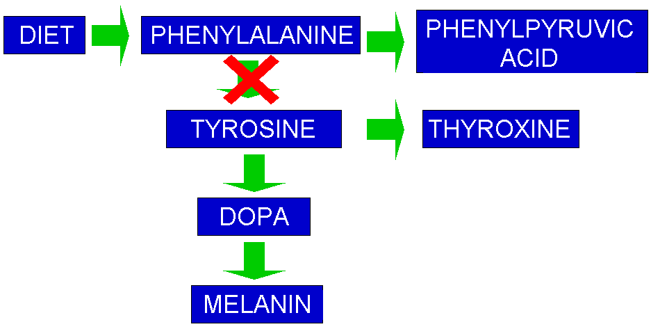
Because PKU occurs in 1 in 10,000 to 15,000 newborns, most babies in the US are screened for PKU shortly after birth. This is done by pricking the heel or hand of the baby and collecting the drops on a Guthrie Card. The blood spot is placed on a plate with a bacteria that cannot grow without PKU. If no bacteria grows then the child does not have PKU. Simple right? If the test is positive further tests are conducted to find if the level of PKU is connected to PAH or the rare (only 2%) defect in the co-factor (BH4) of PAH. he signs and symptoms of PKU vary from mild to severe. The most severe form of this disorder is known as classic PKU. Infants with classic PKU appear normal until they are a few months old. Without treatment with a special low-phenylalanine diet, these children develop permanent intellectual disability. Seizures, delayed development, behavioral problems, and psychiatric disorders are also common. Untreated individuals may have a musty or mouse-like odor as a side effect of excess phenylalanine in the body. Children with classic PKU tend to have lighter skin and hair than unaffected family members and are also likely to have skin disorders such as eczemaAs PAH normally converts phenylalanine to tyrosine, the block occurs between phenylalanine and tyrosine. As phenylalanine cannot be converted to tyrosine it builds up in the blood, when it reaches high levels it is termed hyperphenylalaninemia. This disrupted pathway interrupts an entire process, shown below.


People who have PKU must eat a protein-free diet, because nearly all proteins contain phenylalanine. Infants are given a special formula without phenylalanine. Older children and adults have to avoid protein-rich foods such as meat, eggs, cheese, and nuts. They must also avoid artificial sweeteners with aspertame, which contains phenylalanine.


This has been a 30 minute blogpost. All about PKU. Enjoy.

1. What enzyme is most commonly defective in people with phenylketonuria?


2. What reaction does this enzyme catalyze? (What is the substrate and what product is produced?)

3. Describe the symptoms of phenylketonuria.

4. What causes the symptoms of PKU, the lack of a substance or the buildup of one?

5. How common is phenylketonuria? How is it treated?

Wednesday, November 10, 2010

Cell Membrane Posters.....and Cystric Fibrosis.

So before I get into this post let me begin with a minor disclaimer: Neither Michael and I are "Artistically Gifted" so our poster will NOT be featured in this post.

So basically last week we got the assignment of drawing a poster on the "Fluid Mosaic Cell Membrane Model".  Sounds like fun right?  Surprisingly.............it was, despite our not using markers and any artistic ability since third grade.

Mosaic Model (not our poster)
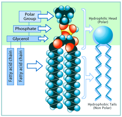
The cell membrane (outside of the cell (works like skin)) is used to control what goes in and out of a cell, ie: food and waste. It is accurately described as a "Fluid Mosaic" because, like glass mosaic it is made up of LOTS of little pieces and like a fluid it is flexible, moving as needed without breaking. The main part of the cell boundary (the "wall" that keeps "stuff" out) is made up of Phospholipids (A hydrophilic phosphorus head (the little circles) attached onto two fatty (lipid) tails). Because the heads are hydrophilic (they like water) they position themselves on the edges of the cell membrane, the hydrophobic ( water fearing- in latin) tails go to the inside of the membrane because they are VERY scared of water. The head of the phospholipid has polar group on the top which forms the bonds that hold mosaic together (from head to head), giving it fluidity.
A Magical Phospholipid!


 Now, do you see those big blue lumbering blobs that are interrupting the beautiful mosaic of phospholipids are proteins, and yes, they serve a function (amazing right?). The middle protein in the picture (the one that looks like it has horns) is called a receptor protein. Receptor proteins are a type of integral protein (proteins integrated into the membrane) that is used for signaling between cells. The receptor proteins absorb the signal molecules sent out by other cells (like in the nerve system) that tell the receiving cell to "fire".  The little valley on the top of that protein is where the molecules "dock". Way out in the back of the protein (back in the middle of nowhere, like La Junta) is a glycoprotein. Glycoproteins are polypeptides (short polymers of amino acids linked by peptide bonds) with a Glycan (the carb structure sticking out) attached. Glycoproteins are used for cell to cell interactions. On the left side of the image there is a recognition protein. Recognition proteins (like their name suggests) are used by cells to identify that they belong in the body. When a person gets an organ transplant the recognition proteins are different so the body will sometimes attack the new organ because it is viewed as a foreign and possibly hostile object.


The last type of protein in the cell membrane is called a transport protein. This protein is possibly the most important protein in the entire membrane (at least in my opinion). Since the membrane (also called the lipid bilayer), is basically a giant impenetrable wall that surounds the yummy goodness that is the internal components of the cell, there needs to be a way to get things in (food, vital molecules) and let stuff (Waste, excess molecules) out. Thus, the transport protein was born. All that a transport protein is simply put, is a doorway into a cell. There is one minor condition though to transport proteins, each one only lets in a certain type of molecule. If I was to use a metaphor to describe it I would compare them to bathroom doors. Only guys go into the men's bathroom and only girls go into the women's bathroom (hopefully). If one sex was to go through the wrong door  (very rare) bad things would happen (think pepper spray and screaming. followed by sexual harassment charges....) Since we have determined that there are specific transport proteins for the different types of molecules (sodium and the like) we now have to think about the two types of transport proteins, defined by how they work.

Transport Proteins.
The first type of transport proteins are channel proteins. Channel proteins span the entire width of the lipid bilayer and use passive transport to move molecules. Passive transport uses no chemical energy to move molecules so it is limited by the size of the molecule. Passive transport is also limited by the chemical gradient (molecules move from high to low concentration).  On a cell membrane this is known as facilitated diffusion.
Facilitated Diffusion
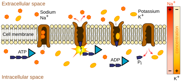
The second type of transport proteins are Carrier proteins. Carrier proteins use chemical energy to move molecules against the concentration gradient and molecules that are to large for a protein channel. Carrier proteins are used to move vital molecules against the concentration gradient so that the cell can survive. If it uses adenosine triphosphate (ATP) as an energy source then it is termed Primary Active Transport. If it uses an electrochemical gradient the it is Secondary Active Transport. 

Primary Active Transport Using ATP
Since transport proteins are necessary for getting molecules in and out of a cell, when something goes wrong with them it can result in pretty nasty disease. Cystic Fibrosis (CF) is one of those diseases. In the cells of people with CF the epithelial cells in the lungs are poorly permeable to Chloride ions (Cl-). Damaged transport proteins are responsible for this.

Epithelial Cells
The lining cells have channels on their outside surface (on the side of the airway). One of the channels allows sodium ions to flow into the cell and the other controls the passage of chloride ions out of the cell into the mucus on the airway surface. Along with the ion pump, the action of the channels results in an excess of chloride ions in the mucus;  an ionic gradient is set up, with a higher concentration towards the outside. In an attempt to equalize the salt concentrations, water is dragged out through the gaps between the cells and this keeps the mucus moist. If a person has CF then the all important chloride channel  is blocked. This means that there is no movement of chloride ions into the mucus. With no ionic gradient, there is no need for water to move towards the surface and the mucus dries out. In 1989 the defective gene for CF was isolated (kinda). There are over 800 different mutations of the gene, with each one affecting the correct function of the Chloride Channel (CF transmembrane regulator (CTFR))  According to <http://resources.schoolscience.co.uk/MRC/3/page3.html> it was 'one of the most significant discoveries in the history of human genetics'. It led directly to improved diagnosis of the disorder and has improved the genetic counseling offered to affected families. 

In conclusion, Holy Crap. These very tiny little cells that make up our body are super complicated and very organized. If one little piece of the cells go bad then your whole body gets screwed up. Its like a swiss watch (had enough of my metaphors yet?) if one little gear is missing it all goes to hell.


Tuesday, November 9, 2010

Carb Concept Map

So I finally finished my concept map about carbs. Granted this was like a month ago but, this time I clicked the right button. So here we go my 1st biochemistry post with one on lipids and fats coming sometime in the next two weeks.

Thursday, October 7, 2010

Research Article......Standard 2!

So basically the first part of this article started out talking about how hard it is to measure "surface coatings of water".  The reason that water is so hard to measure is water molecules are in constant flux. Professor James Heath from CalTech, and his team, have developed a way to trap those water molecules at room temperature, and it was a complete accident. They were studying graphene on an atomically flat surface of mica and found some nanoscale island-shaped structures trapped between the graphene and the mica that they had not expected to see. Graphene is basically an atomic sized chicken wire shaped lattice of carbon molecules. Heath and his team thought that the bumps might be water, it is everywhere.... After discovering this they decided, being scientists and all, to test it. After varying the humidity in the room, high to low, they observed that the bumps disappeared when humidity was low, and reappeared when the humidity was high. They then realized that the graphene was basically "atomic shrink wrap" once the molecules were trapped they could probe the molecules with an Atomic Force Microscope. Using this method the researchers discovered new details on how water coats ice! What they discovered is that the first layer of water is two molecules thick, and has the structure of Ice. Once that layer is fully formed another layer of two molecule thick ice is added, which is then topped by ice. These newly discovered structures are likely important in determining the surface properties of solids, like: lubrication, adhesion, and corrosion. This technique could later be used to make 3-D images of molecules. This may make it possible to analyze very complex molecules like protien-protein complexes, revealing ever single little nook and cranny. Being able to get pictures of these molecules could prove very important to how molecules are understood.


Water trapped under the Graphene

http://www.futurity.org/science-technology/dead-simple-way-to-see-atomic-structure/

Tuesday, September 21, 2010

Reflective Post....on pH stuff...whoo...

So pH levels....pretty cool stuff right? pH levels are determined by the concentration of ions in a substance. In pure water (H2O), there is 1 ion for every 555 million water molocules, this can be noted as 1x10^-7. On the pH scale water is a 7 (neutral). The number on the pH scale is the exponent on the scientific notation (not the log... Michael) of the concentration of hydrogen ions. Acids are H+, meaning they have a higher concentration of Hydrogen ions which makes the exponent smaller (1x10^-2, or something......you do know what an exponent is right?) Bases, which are on the other end of the pH scale have more OH-. Since the have more OH- their concentration of H+ is smaller, resulting in a higher exponent value(seems backwards right?).   Water.....the magical liquid of life has a pH level of 7. pH level 7 is in the middle of the pH table (0-14....do the math). Water is special enough to get the pH level of 7 because it has one H+ and one OH- ion. (H+) + (OH-) = (H2O). Since water has one of each charged molocule it is neutral....a positive and a negative charge cancel out.

Monday, September 20, 2010

Acids In Your Tummy Log.....Whoo!!!

Here is the write up for the Lab on the effectiveness of different antacids. Contributors Include Michael Rees, Tyler White, Seth Nixon, and Myself.

Friday, September 3, 2010

Clinical Trials.....

So this week we have been working with clinical trials in Biology. Go Experimental Drugs! Since we are graded on proving that we actually learned something this week rather than just screwing around on the internet, I decided to write a post about what I learned, so here goes.....

Once upon a time in a world far far away, there was a brilliant scientist named Flinn who had just discovered an amazing new drug that seemed to cure severe lung cancer. In another part of the world a small child named Bob (lame right?) who had just been diagnosed with lung cancer, and had been given only 4 months to live because there was no cure for the cancer. Now Bob's doctor had just read an article about Flinn's drug and knew that it was entering into the clinical trial phase. Thinking that this was Bob's best hope Greg (the doctor) got Bob enrolled in the trial (Yay!). Now the trial itself was a Double Blind Placebo meaning that patients that are receiving the drugs don't know if they are taking the placebo (fake drug with no effect) or the experimental drug. In a double blind study the staff that hand out the drugs and record the changes also don't know what drugs the patients are taking. This type of procedure helps prevents false and bias results in clinical trails. Now little Bob got extremely lucky because the drug worked and cured the cancer. And they all lived happily ever after.